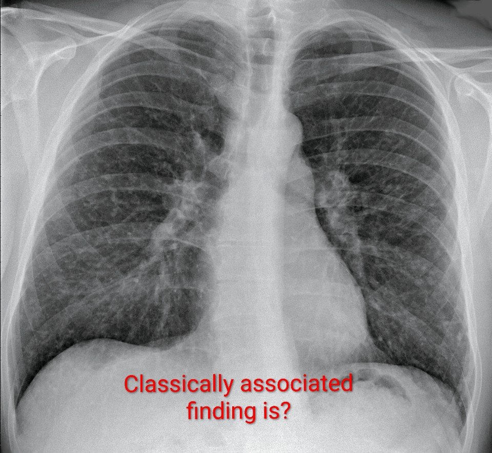
Q. A 45-year-old man with sarcoidosis has a chest X-ray showing bilateral perilymphatic pulmonary nodules. Which associated finding is classically expected?
A) Pleural effusion
B) Lobar consolidation
C) Cavitary lesions
D) Bilateral hilar lymphadenopathy
✓ The Correct Answer is: D) Bilateral Hilar Lymphadenopathy.
High-Yield Review
Sarcoidosis is a multisystem granulomatous disorder characterized by the formation of non-caseating granulomas. The lungs and intrathoracic lymph nodes are most commonly involved.
A classic chest-imaging pattern is the combination of bilateral hilar lymphadenopathy with perilymphatic pulmonary nodules.
The nodules tend to follow the lymphatic pathways, particularly along the bronchovascular bundles, interlobular septa, and subpleural regions.
This makes bilateral hilar lymphadenopathy a key imaging clue in sarcoidosis.
Why Does Hilar Lymphadenopathy Occur?
Sarcoidosis causes granulomatous inflammation within the lymphatic system.
This results in enlargement of the bilateral hilar and mediastinal lymph nodes.
On chest imaging, the symmetrical hilar enlargement can produce the classic “potato nodes” appearance.
The pulmonary nodules, meanwhile, reflect granulomatous involvement along the pulmonary lymphatic routes.
Other Chest X-Ray Findings
Depending on the stage and severity, sarcoidosis may show:
Bilateral hilar lymphadenopathy.
Mediastinal lymphadenopathy.
Perilymphatic pulmonary nodules.
Upper-lobe predominant reticulonodular opacities.
Fibrotic changes in advanced disease.
Architectural distortion and volume loss in late-stage disease.
∆ Other Options:
Pleural effusion: It is not a typical feature of sarcoidosis and should prompt consideration of another cause.
Lobar consolidation: It is more suggestive of infection, pulmonary edema, or other focal air-space disease.
Cavitary lesions: They are uncommon in sarcoidosis and are not a classic radiological finding.
Bilateral hilar lymphadenopathy: It is classic and strongly associated with pulmonary sarcoidosis.
∆ High-Yield Exam Pearls:
Sarcoidosis → Think:
Non-caseating granulomas.
Bilateral hilar lymphadenopathy.
Perilymphatic pulmonary nodules
Upper-lobe predominant involvement.
ACE may be elevated but is neither sufficiently sensitive nor specific to establish the diagnosis.
Löfgren syndrome: Erythema nodosum + bilateral hilar lymphadenopathy + arthritis/arthralgia, often involving the ankles.
✓ One-Liner to Remember:
“Perilymphatic nodules + bilateral hilar lymphadenopathy = classic pulmonary sarcoidosis.”
✓ Take-Home Message:
In a patient with sarcoidosis, bilateral hilar lymphadenopathy is the classic associated chest-imaging finding when perilymphatic pulmonary nodules are present.
Recognizing this combination is particularly useful for image-based questions and clinical practice and is a high-yield concept for MBBS, NEET PG, INICET, FMGE, USMLE, and PLAB examinations.