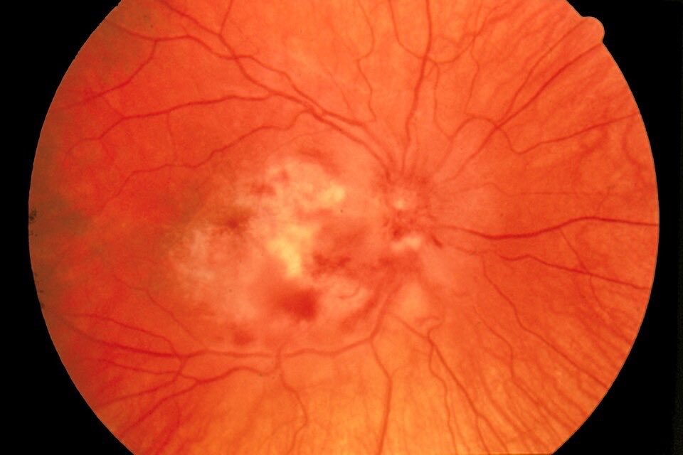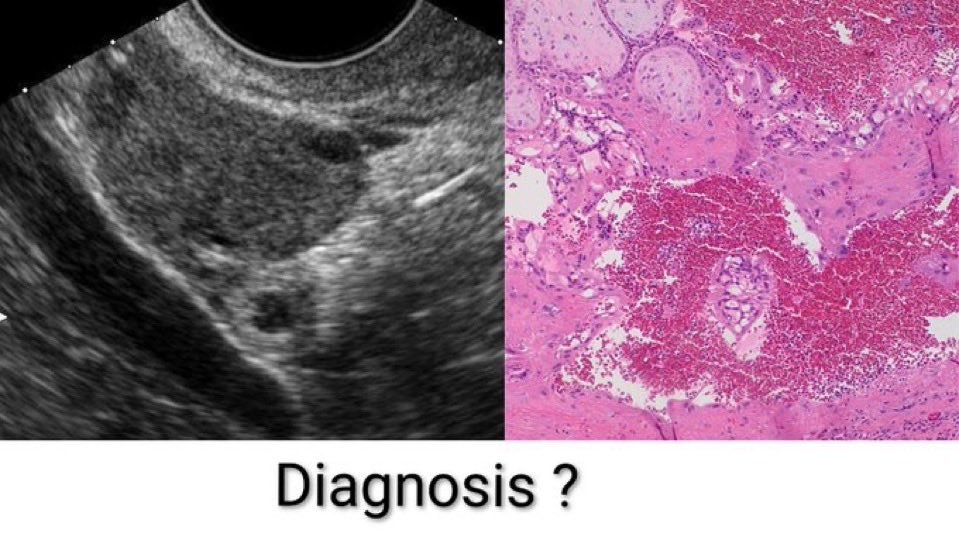
Q. A 25-year-old woman presents with 6 weeks of amenorrhea, mild lower abdominal pain, and spotting per vaginum. Her urine pregnancy test is positive. Ultrasound & Histopathology images are shared above. What is the most likely diagnosis?
A) Incomplete abortion.
B) Ectopic pregnancy.
C) Hydatidiform mole.
D) Corpus luteum cyst.
✓ The Correct Answer is: B) Ectopic Pregnancy.
A woman of reproductive age presenting with amenorrhea, abdominal pain, and vaginal spotting should always raise immediate suspicion for an ectopic pregnancy until proven otherwise.
An ectopic pregnancy occurs when the fertilized ovum implants outside the uterine cavity, with the fallopian tube (especially the ampulla) being the most common site.
As the pregnancy grows, it stretches the surrounding tissue, causing pain and bleeding. If not diagnosed early, tubal rupture can lead to life-threatening intra-abdominal hemorrhage.
∆ Clinical Features
The typical presentation includes-:
1. 6–8 weeks of amenorrhea.
2. Positive urine or serum pregnancy test.
3. Lower abdominal or pelvic pain.
4. Light vaginal bleeding or spotting.
5. Adnexal tenderness or cervical motion tenderness on examination.
6. In cases of rupture, patients may develop severe abdominal pain, dizziness, hypotension, tachycardia, and signs of hemorrhagic shock, requiring immediate intervention.
∆ Diagnosis
Transvaginal ultrasonography combined with serial serum β-hCG measurements forms the cornerstone of diagnosis.
A positive pregnancy test with no intrauterine gestational sac visible on transvaginal ultrasound when the β-hCG level is above the discriminatory zone strongly suggests an ectopic pregnancy.
Ultrasound may also reveal an adnexal mass or free fluid in the pelvis, indicating bleeding.
∆ Other Options-:
Incomplete abortion usually presents with an open cervical os and retained products of conception within the uterus.
Hydatidiform mole is characterized by markedly elevated β-hCG levels, excessive uterine enlargement, and the classic "snowstorm" appearance on ultrasound.
Corpus luteum cyst may cause pelvic pain but does not typically present with the classic triad of amenorrhea, positive pregnancy test, and vaginal spotting due to an extrauterine pregnancy.
∆ High-Yield Exam Pearls:
Most common site of ectopic pregnancy: Ampulla of the fallopian tube.
Most common risk factor: Previous tubal damage (e.g., pelvic inflammatory disease or prior ectopic pregnancy).
Investigation of choice in a stable patient: Transvaginal ultrasound with quantitative serum β-hCG.
Medical management: Methotrexate in carefully selected stable patients.
Emergency treatment: Laparoscopic surgery for ruptured or unstable ectopic pregnancy.
✓ One-Liner to Remember:
"Amenorrhea + abdominal pain + vaginal spotting + positive pregnancy test = Think ectopic pregnancy until proven otherwise."
✓ Take-Home Message:
Ectopic pregnancy is a time-sensitive obstetric emergency that requires early recognition and prompt treatment to prevent maternal morbidity and mortality.
The combination of amenorrhea, lower abdominal pain, vaginal spotting, and a positive pregnancy test should immediately prompt evaluation with transvaginal ultrasonography and serum β-hCG.
As this clinical scenario is repeatedly tested in MBBS, NEET PG, INICET, FMGE, PLAB and USMLE examinations, every medical student should be able to identify it quickly and confidently and this will be quite useful in clinical practice as well.